Health Articles
We Got Your Back


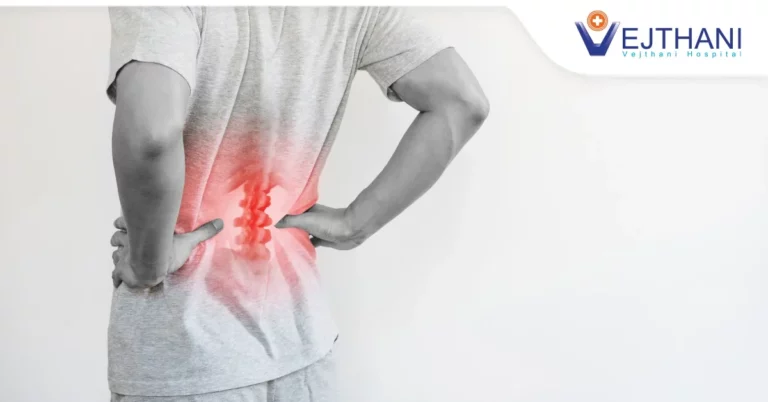
Your posture affects your physical appearance the most. Starting from the way you stand up to the fine movements you could do every day. Imagine not standing with good posture and a straight back, then talking to somebody; let’s say your client or even your friend, and most especially speaking or walking in public. It would be very disturbing and could affect our self-esteem. Likewise, when doing your usual household chores, it should be done with good posture. All of these activities could be affected if your spinal column, particularly your lumbar spine (lower back) is damaged due to certain diseases or accident.
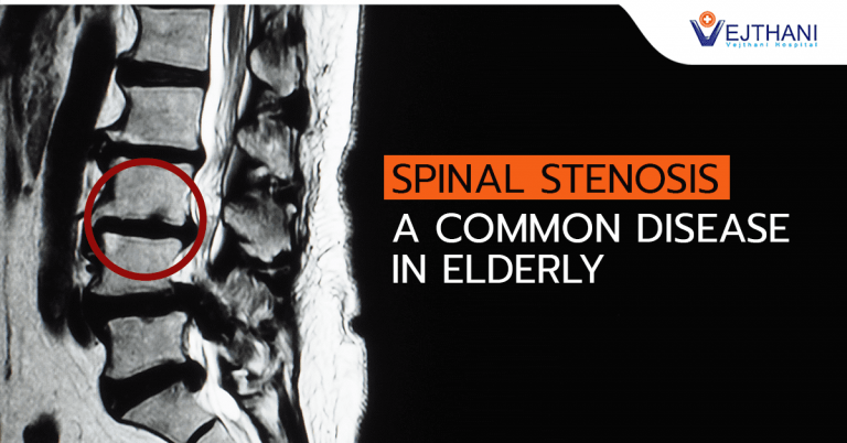
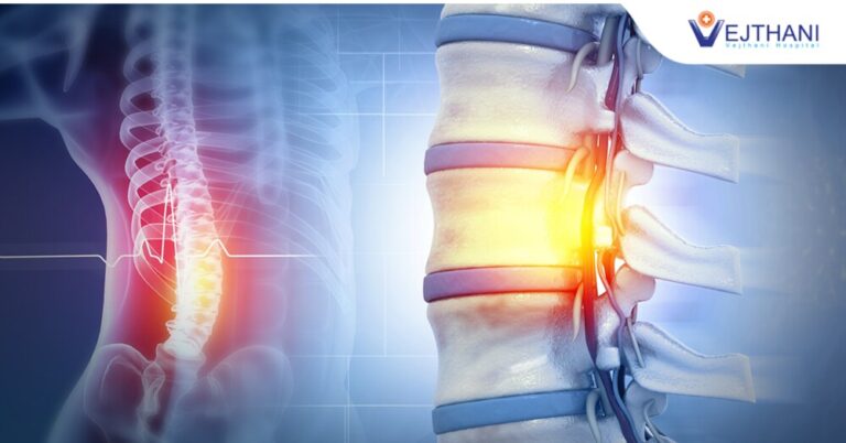
The lumbar vertebrae consist of five individual cylindrical bones that form the spine in the lower back. They are the largest segments of the vertebral column. It is built for both power and flexibility – lifting, twisting, and bending. They also protect the delicate spinal cord and nerves within their vertebral canal. Therefore, if the lumbar spine is damaged, no more lifting, twisting, and bending activities.
XLIF (Extreme Lateral Interbody Fusion)
The XLIF (eXtreme Lateral Interbody Fusion) is a minimally invasive approach to spinal fusion in which the surgeon accesses the intervertebral disc space and fuses the lumbar spine (low back) using a surgical approach from the side (lateral) rather than from the front (anterior) or the back (posterior). Instead of a traditional, larger single incision, the procedure is performed through one or more small incisions and an instrument known as a retractor is used to spread the tissues so that the surgeon can see the spine. However, it cannot be used for all types of lumbar conditions for which spinal fusion is a treatment option. For example, it cannot treat conditions at the lowest level of the spine, L5-S1 or for some people at L4-L5.
Indications
XLIF treats specific types of lumbar spinal disorders, such as:
- Degenerative disc disease
- Spondylolisthesis,
- Scoliosis and deformity and some types of lumbar stenosis
- Recurrent lumbar disc herniations
- Foraminal stenosis (a type of spinal stenosis)
- Thoracic disc herniation
Advantages
- Minimal tissue damage
- Minimal blood loss
- Small incisions and scars
- Minimal post-operative discomfort
- Relatively quick recovery time and return to normal function.
- Does not require dissection or retraction of the sensitive back muscles, bone, or never,
- Nor does it require the delicate abdominal exposure
How is the procedure done?
- First, the patient will be positioned lying on his or her side. Then the surgeon will use X-rays to locate the disc that will be removed.
- Once the disc is located, the surgeon will mark the skin with a marker directly above the disc.
- Then the surgeon will make a small incision (cut) in the flank (low back region of the trunk) and use his or her finger to push away the peritoneum (sac covering the abdominal organs) from the abdominal wall.
- The surgeon will make a second incision directly on the side of the patient.
- The surgeon will then insert a tube-like instrument known as a dilator into this incision.
- The surgeon will use X-rays to make sure that this dilator is in a good position above the disc.
- The surgeon will then insert a probe (blunt tool) through a muscle known as the psoas muscle. The psoas muscle is a large muscle that runs from the lower spine, wrapping around the pelvic area and attaches at the hip.
Because nerves exiting the spinal column are close to the psoas muscle and can even run right over the surface of it, it is critical that the surgeon be provided with real-time information about the position of the nerves relative to his instruments. Neuromonitoring, the testing of the nerves during surgery to make sure that they are not harmed or irritated during the process, is a critical part of this procedure. This type of nerve monitoring is known as electromyography or EMG. The neuromonitoring system used with the XLIF procedure is designed to accomplish this goal. The probe is designed to detect the position of the nerves to help avoid disturbing them during surgery.
- Once the muscles are split apart, a retractor is put into place to give the surgeon direct access to the spine.
- Once this direct access to the spine is achieved, the surgeon is able to perform a standard discectomy (removing the intervertebral disc) with tools designed to cut and remove the disc.
- After the disc material is removed, the surgeon is able to insert the implant through the same incision from the side. This spacer (cage) will help hold the vertebrae in the proper position to make sure that the disc height (space between adjacent vertebral bodies) is correct and to make sure the spine is properly aligned. This spacer (together with the bone graft, is designed to set up an optimal environment to allow the spine to fuse at that particular segment.
- The surgeon will take an X-ray to make sure that the spacer is in the right position.
Recovery Period
- The total time a patient spends in the hospital after the surgery depends on several factors, such as the number of vertebral levels that were fused, the severity of the problem and the patient’s overall health.
- Some patients who undergo an XLIF procedure are able to return home the same day as the surgery; others require a stay of a few days or a week in the hospital.
- Most patients are able to return to their normal activities within a few months of surgery.
Vejthani Hospital Spine Center offers a wide range of premium services including consultation and diagnosis, treatment, rehabilitation, and physiotherapy. Vejthani Hospital Spine Center makes use of only the latest hi-technology medical facilities such as CT scan and the like. All is to guarantee our patients to receive the finest treatment in spine surgery right here in Vejthani Hospital.
At Vejthani Hospital Spine Center, we place a strong emphasis on individualized care with treatment options designed to meet each patient’s specific needs and goals. We provide a team approach to rehabilitative care, neck and spine health and pain management to restore our patients to their most productive and best health.
You will be cared for by a team of experts providing the most advanced treatment options and comprehensive care available to prevent, treat and rehabilitate conditions that affect the spinal column. Most importantly, this combined advanced medicine is tailored to your individual needs.
For inquiries or appointment Vejthani Hospital Spine Center is located at fifth floor or contact us through: email int_mkt@www.vejthani.com
Tel +66 (0) 2734-0000 ext 5500/5550
Fax +66 (0) 2734-0375.
Service hours starts 8:00 am up to 8:00 pm from Monday to Sunday.
