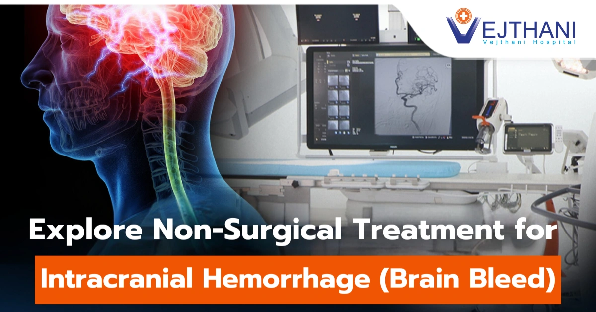
Arteriovenous malformations
Overview
Arteriovenous malformations (AVM) are an abnormal tangle of blood vessel that connect arteries and veins and impair normal blood flow and oxygen delivery. Although they can arise anywhere in your body, they mostly happen in your brain and spinal cord. Blood carrying oxygen from the heart to the brain is carried via arteries. The oxygen-depleted blood is then returned to the heart and lungs through veins.
The surrounding tissues may not receive adequate oxygen when an AVM interferes with this crucial process. Additionally, the abnormally tangled blood arteries that make up the AVM are prone to being damage and rupture, due to the pressure brought by the force of the blood flow. If the AVM ruptures in the brain, it may result in bleeding within brain (hemorrhage), which results in a stroke, or permanent brain damage.
AVMs have an unknown cause. They are rarely passed down among families. Once identified, a brain AVM can frequently be successfully treated to prevent complication.
Symptoms
Symptoms of an AVM may vary depending on where it is located. The most common site of AVM is found in the brain, which could results in brain hemorrhage. As a result of the surrounding tissue being interfere by AVMs, neurologic symptoms such as the following occur:
Brain AVM:
- Seizures
- Loss of consciousness.
- Headache, or dizziness.
- Muscle weakness, numbness, or tingling sensation.
- Paralysis.
- Nausea and vomiting.
- Impairment of balance, vision, memory and speech
- Mental confusion, hallucinations or dementia.
Spinal cord AVM:
- Back discomfort (abrupt onset and severe pain)
- Weakness in your legs, hips, and toes.
- Muscle weakness or paralysis due to affected nerves.
AVMs in other parts of the body: Depending on their size and the significance of the location, AVMs in other parts of the body (other than the brain and spine) may or may not cause symptoms. Typical general signs include:
- Shortness of breath or difficulty in breathing.
- Coughing up blood
- Abdominal pain.
- Lumps on the arms, legs, or trunk.
- Pain and swelling.
- Muscle weakness or paralysis.
- Tingling sensation or numbness.
- Open ulcers at the skin.
Congenital AVM: Symptoms of one type of AVM known as a vein of Galen defect which is a defect deep in the brain, start to show up at or soon after birth. An example of a sign is:
- Hydrocephalus: enlargement of the head due to fluid accumulate in the brain’s ventricles
- Swollen veins on the scalp
- Seizures
- Failure to thrive
- Congestive heart failure
If you have any of these symptoms, seek emergency medical attention immediately:
- Severe headaches
- Vision problems
- Seizure
- Weakness
- Changes in behavior or neurological function
Causes
AVMs develop when arteries and veins develop abnormal connections, although researchers are unsure what cause it to happen. Though most types are typically not inherited, some genetic modifications might be involved.
Risk factors
The following increases the risk to develop AVM are:
- Family history: At rare cases, it increases the risk to develop AVM if there are history of the condition within the family. However, most cases of AVM are not inherited. (1)
- Hereditary conditions: risk of AVM may be increased by some inherited disorders. These include Osler-Weber-Rendu syndrome and hereditary hemorrhagic telangiectasia (HHT).


