Stroke
Diagnosis
Diagnosing a stroke requires a healthcare provider to employ a combination of methods, including a neurological examination, diagnostic imaging, and other tests. During the neurological examination, the provider will ask you to perform specific tasks or answer questions. By observing your performance and responses, the provider can identify characteristic indicators that suggest dysfunction in a particular area of your brain. When a healthcare provider suspects a stroke, several commonly conducted tests are typically performed that including:
- Blood tests. Several blood tests may be conducted as part of the medical evaluation. These tests can assess various factors such as blood clotting speed, blood sugar levels, and the presence of any infections.
- Computerized Tomography (CT) scan. An accurate image of your brain is produced by a CT scan using a sequence of X–rays. A CT scan can detect tumors, ischemic strokes, brain tumors, and other disorders. To observe the blood vessels in the neck and brain in greater detail using computed tomography angiography, doctors may inject a dye into your circulation.
- Magnetic Resonance Imaging (MRI). A magnetic field and strong radio waves are used in an MRI to produce a precise image of the brain. Brain hemorrhages and ischemic stroke damage can both be found on an MRI. To view the arteries and veins and highlight blood flow, your doctor may inject a dye into a blood vessel (magnetic resonance angiography or magnetic resonance venography).
- Carotid ultrasound. The carotid arteries in the neck are visualized inside this test using finely detailed sound waves. Performing this test reveals blood flow in the carotid arteries as well as plaque buildup.
- Cerebral angiogram. In this infrequently performed test, your doctor makes a tiny incision, typically in the groin, and inserts a thin, flexible tube (catheter), guiding it through the major arteries and into the carotid or vertebral artery. The blood vessels are then given a dye injection by your doctor to make them visible on an X–ray. The arteries in the brain and neck can be seen in great detail thanks to this treatment.
- Echocardiogram. Sound waves are used in an echocardiography to provide fine–grained pictures of the heart. An echocardiography can identify the origin of any cardiac clots that may have caused a stroke by moving from the heart to the brain.
- Electroencephalogram (EEG). While less common, an EEG can be conducted by a healthcare provider to rule out seizures or related issues.
Treatment
Your healthcare provider will develop a personalized care plan considering various factors:
- Age, overall health, and medical history: Your age, general health condition, and past medical experiences will be taken into account to determine the most suitable approach.
- Type and severity of stroke: The specific type of stroke you experienced (e.g., ischemic or hemorrhagic) and its severity will be considered in creating your care plan.
- Location of the stroke in the brain: The area of the brain affected by the stroke will influence the recommended treatments and therapies.
- Stroke cause: Understanding the underlying cause of your stroke (e.g., blood clot, high blood pressure, or other factors) is essential for developing an appropriate care plan.
- Response to medications, treatments, and therapies: Your healthcare provider will assess how well you respond to different medications, treatments, or therapies to determine the most effective options for you.
While stroke cannot be cured once it has occurred, advancements in medical and surgical interventions can help lower the risk of future strokes. These interventions will be considered in your care plan to provide the best possible outcomes.
Ischemic stroke
Blood flow to the brain needs to be immediately restored for an ischemic stroke to be treated. This is possible by using:
- Emergency IV medication. Therapy involving the use of clot–dissolving drugs needs to be administered intravenously within 4.5 hours from the onset of stroke symptoms. The earlier these medications are given, the greater the chances of survival and the potential reduction of complications. The preferred treatment for ischemic stroke is an intravenous injection of recombinant tissue plasminogen activator (TPA), which is also known as alteplase (Activase) or tenecteplase (TNKase). Typically, TPA is administered through a vein in the arm within the first three hours after stroke symptoms appear. In some cases, TPA can be given up to 4.5 hours after symptom onset. This medication works by restoring blood flow through the dissolution of the blood clot responsible for the stroke. By swiftly addressing the underlying cause of the stroke, TPA may aid in a more complete recovery. Your doctor will assess certain risks, such as the possibility of bleeding in the brain, to determine the appropriateness of TPA treatment for you.
Emergency endovascular procedures are employed by doctors to directly address ischemic strokes occurring within blocked blood vessels. These procedures, known as endovascular therapy, have demonstrated significant improvements in outcomes and reduction of long–term disability following ischemic stroke. It is crucial that these procedures are performed promptly. There are two primary techniques utilized:
- Direct delivery of medications to the brain: To administer treatment directly at the site of the stroke, doctors insert a long, thin tube called a catheter through an artery in the groin. The catheter is then guided to the brain, allowing for the delivery of medication such as tissue plasminogen activator (TPA). This method has a slightly longer time window for treatment compared to injected TPA, but it remains time–limited.
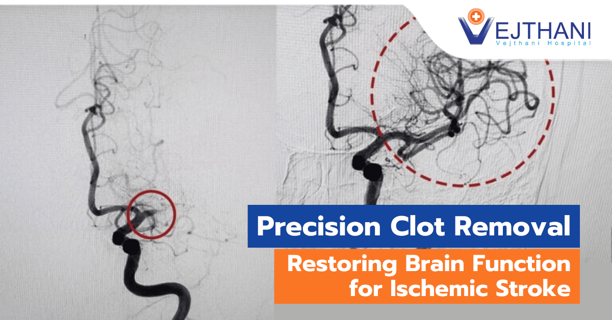
- Clot removal using a stent retriever: In cases where large clots are present and cannot be fully dissolved with TPA, doctors can employ a device attached to a catheter to physically remove the clot from the blocked blood vessel within the brain. This procedure is often performed alongside the administration of injected TPA, maximizing the effectiveness of treatment.
Other procedures
Your doctor might advise an operation to widen an artery that has been constricted by plaque in order to reduce your risk of experiencing another stroke or transient ischemic attack.
Depending on the circumstance, options can include:
- Carotid endarterectomy. The carotid arteries are the blood vessels that run down each side of the neck and carry blood to the brain. By removing the plaque that is obstructing a carotid artery, this procedure may lower the risk of an ischemic stroke. Additionally risky is a carotid endarterectomy, particularly for those with heart disease or other medical disorders.
- Angioplasty and stents. A catheter is inserted into the carotid arteries during an angioplasty through a groin artery. The artery is then widened by inflating a balloon. The opened artery can then be supported by the placement of a stent.
Hemorrhagic stroke
The primary goals of emergency hemorrhagic stroke treatment are to stop the bleeding and ease the pressure that the extra fluid is putting on the brain. Options for treatment include:
- Emergency treatment. You may receive medications or blood product infusions to offset the effects of blood thinners if you use them to avoid blood clots. Additionally, medications may be administered to you to lower blood pressure, stop blood vessel spasms, stop seizures, and lower intracranial pressure.
- Surgery. Your doctor may conduct surgery to drain the blood and relieve pressure on the brain if the bleeding is extensive. Additionally, surgery may be performed to treat blood vessel issues brought on by hemorrhagic strokes. After a stroke or if an aneurysm, arteriovenous malformation (AVM), or other blood vessel issue caused the hemorrhagic stroke, your doctor may advise one of these operations.
- Surgical clipping. In order to stop blood flow to the aneurysm, a surgeon applies a small clamp on its base. This clamp can prevent an aneurysm from rupturing or prevent a recently hemorrhaged aneurysm from bleeding again.
- Coiling (endovascular embolization). The surgeon will inject tiny detachable coils into the aneurysm to fill it using a catheter that is placed into an artery in the groin and directed to the brain. As a result, the aneurysm is unable to receive blood flow and clots.
- Removing AVM by surgery. A minor AVM may be removed by surgery if it is found in a region of the brain that is easily accessible. This reduces the risk of hemorrhagic stroke and removes the possibility of rupture. An AVM may not always be able to be removed if it is large, deeply positioned in the brain, or if doing so would have a negative impact on brain function.
- Stereotactic radiosurgery. Stereotactic radiosurgery is a sophisticated minimally invasive procedure used to treat blood vessel abnormalities. It uses multiple beams of highly concentrated radiation.
Stroke recovery and rehabilitation
After receiving emergency treatment for a stroke, the focus of care shifts towards aiding the recovery process and helping the individual regain as much functionality as possible in order to return to independent living. The extent of the stroke’s impact varies based on the specific region of the brain affected and the amount of tissue damage incurred. If the right side of the brain is affected, it may lead to impaired movement and sensation on the left side of the body. Conversely, damage to the left side of the brain can result in compromised movement and sensation on the right side, as well as speech and language disorders.
Rehabilitation programs play a crucial role in the recovery of stroke survivors. The chosen program will depend on factors such as the person’s age, overall health, and the degree of disability caused by the stroke. Additionally, the individual’s lifestyle, interests, and availability of family members or caregivers are taken into consideration. Rehabilitation may commence during the hospital stay and can continue in various settings, including specialized units within the hospital, other rehabilitation facilities, outpatient settings, or even at home.
The stroke recovery journey is unique to each individual, and a comprehensive treatment team is usually involved. This team may consist of medical professionals such as neurologists and physiatrists, as well as rehabilitation nurses, dietitians, physical therapists, occupational therapists, recreational therapists, speech pathologists, social workers or case managers, psychologists or psychiatrists, and chaplains. The collective expertise of these professionals helps address the diverse needs of stroke survivors, enabling them to achieve the best possible outcomes and improve their quality of life.


