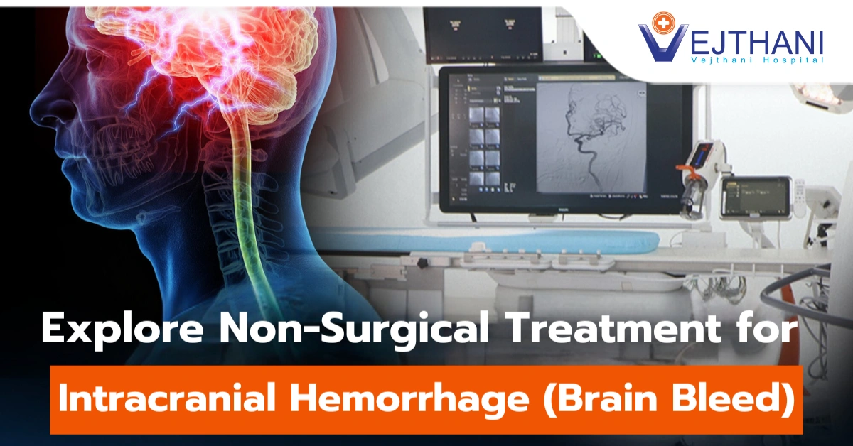
Gamma knife radiosurgery, Gamma brain stereotactic radiosurgery, gamma knife radiation
Overview
Gamma Knife radiosurgery is a non-invasive form of radiation therapy designed to treat conditions affecting the brain and upper spine. This technique employs computer-guided planning and can effectively address tumors, abnormal blood vessels, and other brain abnormalities.
Unlike traditional surgery, Gamma Knife radiosurgery does not involve any incisions. It precisely targets specific areas of the brain without the need to penetrate the skin. By utilizing multiple tiny radiation beams, this procedure delivers a highly focused dose of radiation to the tumor or targeted area while minimizing damage to surrounding healthy tissue. The convergence point of these beams receives a concentrated dose of radiation.
Also referred to as Gamma brain stereotactic radiosurgery or Gamma Knife radiation, this treatment is typically administered once a day.
Reasons for undergoing the procedure
Compared to standard brain surgery, Gamma Knife radiosurgery can be safer. Cutting into brain tissue and creating incisions in the head, skull, and membranes around the brain are all part of standard surgery. Typically, this kind of radiation therapy is carried out when:
- Standard neurosurgery cannot reach a tumor or other abnormality in the brain.
- The patient’s condition isn’t suitable for standard surgery.
- A less invasive course of treatment is preferred.
When compared to other forms of radiation therapy, Gamma Knife radiosurgery often has fewer side effects. This type of procedure just takes a single day, while standard radiation therapy may require up to thirty treatments.
Gamma Knife surgery is intended to either shrink, destroy, or stabilize a tumor or lesion. Treatments for the following conditions are most frequently treated with gamma knife radiosurgery:
- Acoustic neuroma/ vesticular schwannoma: A noncancerous growth is called an acoustic neuroma. You may have tinnitus, or ringing in the ears, as well as hearing loss, vertigo, and loss of balance when the tumor presses and put pressure against the nerve. The nerves that regulate facial muscles and feelings may also be compressed by the growing tumor. An acoustic neuroma may not grow further if radiosurgery is performed.
- Arteriovenous malformation (AVM): The blood arteries in the AVM eventually close as a result of radiosurgery. The chance of bleeding is decreased by this procedure.
- Brain tumor: Small benign (noncancerous) brain tumors can be treated with radiosurgery. Malignant brain tumors can also be treated with radiosurgery.
The genetic substance in tumor cells known as DNA is damaged by radiosurgery. The tumor may progressively shrink as a result of the cells’ inability to proliferate and may eventually die.
- Pituitary tumors: Pituitary hormone secretion can be reduced and the tumor can decrease with radiosurgery.
- Trigeminal neuralgia: The brain and regions of the forehead, cheek, and lower jaw communicate with each other through the trigeminal nerves. Facial discomfort associated with trigeminal neuralgia has an electric shock-like sensation. Pain reduction with treatment may occur in a few days to several months.
Among the most common conditions that are treated using Gamma Knife surgery are:
- Arteriovenous malformations (AVM).
- Epilepsy.
- Essential tremor or Parkinson’s disease symptoms.
- Trigeminal neuralgia.
Risk
Compared with standard neurosurgery, gamma knife radiosurgery is typically low risk because it doesn’t require any surgical incisions.
Early side effects or risks are typically temporary. The following includes:
- A tingling or numb feeling at the pin placement areas on the scalp.
- Brain swelling usually show up about six months after treatment rather than immediately after the procedure like with standard surgery.
- Fatigue at the first few weeks after the procedure.
- Headache.
- Nausea and vomiting.
- Scalp and hair issues: In rare cases, if the area being treated is directly beneath the scalp, some patients may experience a temporary loss of hair.
- Skin discoloration and bruises where pins are placed.
Before the procedure
A consultation with a healthcare provider will take place before the Gamma Knife procedure. This healthcare provider may be a specialist of radiation oncology or a neurosurgery. They will examine you physically first. Inform your healthcare provider about any medical implants you may have during this examination, such as a cochlear implant, pacemaker, implanted medication pump, nerve stimulator, or any other type of implant.
In order to complete the process, they will also need to know if you can lie flat on your back for thirty to sixty minutes. If you have reflux or lung disease, this may be challenging for you to perform.
Particular preparation instructions for your Gamma Knife surgery will be given to you. These instructions may, in general, consist of:
- Avoid eating or drinking anything after midnight before your procedure.
- Drink little amounts of water when taking your morning meds. Remember to pack all of your prescription and over-the-counter (OTC) drugs, including inhalers.
- The night before your Gamma Knife surgery, wash your scalp. Let your hair hang loose. Avoid wearing bands, clips, or pins in your hair the day of the operation.
- Wear loose-fitting, comfy clothing and shoes that are simple to take off. Stay clear of putting on or taking off shirts over your head.
- You will require a ride to and from the procedure from someone.
Four pins will be used to secure a thin frame to your head prior to the procedure begin. Your head will remain steady with this frame while receiving radiation therapy. The radiation beams will be focused utilizing the frame as a point of reference. Throughout this procedure: (1 all)
- You won’t have your hair shaved, but rubbing alcohol will be used to clean your forehead and back of your head.
- The four areas of your scalp where the pins will be inserted—two on each forehead and two at the rear of the head—will be numbed with injections.
Following the attachment of the head frame, brain imaging is carried out to determine the tumors or other target’s location in relation to the head frame. The condition that is being treated determines the type of scan that is used:
- Tumors: Magnetic resonance imaging (MRI) or computed tomography (CT) can be used for tumor imaging. A CT scan uses a series of X-rays to produce a comprehensive picture of the brain. A magnetic field and radio waves work together to produce precise images of the brain during an MRI exam.
In order to inject a dye into a blood vessel, a small needle may be inserted into the arm or the back of the hand. The color makes blood circulation easier to see and makes the blood vessels in the brain easier to see. You could occasionally need both an MRI and a CT scan.
- Arteriovenous malformations (AVMs): brain imaging MRI scans and cerebral angiograms are frequently utilized for AVMs. During a cerebral angiography, a healthcare provider uses X-ray imaging to thread a small tube through a blood vessel in your groin and into your brain. In order to see the blood arteries on an X-ray, dye is injected through them. During CT or MRI scans, your healthcare provider could inject a dye into a blood vessel to observe the blood vessels and emphasize blood circulation.
- Trigeminal neuralgia: To choose a target location for trigeminal neuralgia treatment, an MRI is used to obtain pictures of nerve fibers.
The brain scan data is entered into a computer. The radiosurgery team uses a specific planning program to assist them choose which locations to treat, how much radiation to give, and how to concentrate the radiation beams. Usually, this planning process takes about sixty minutes. You are free to unwind in another room during that period, but the frame needs to stay on your head.
Children are frequently given medications to induce a sedative condition in preparation for radiosurgery and imaging testing. Adults are often awake, however they could receive medication to aid in relaxation.
During the procedure
You will be advised to lie on the bed, and the bed will move into the Gamma Knife machine to deliver the radiation. Your head and body may be slightly moved by the machine to ensure that all treatment areas are exposed to radiation. The helmet inside the machine is firmly attached to the head frame.
To stay hydrated throughout the day, you will get an intravenous (IV) tube that will supply fluids to your bloodstream. The IV’s needle is inserted into a vein, probably in your arm.
The process can take anything from under an hour to over four hours to finish. The target’s size and shape will determine the length. Throughout the process you will not feel the radiation and will not hear any noise from the machine:
Throughout the process, your healthcare team will be just outside the room while you receive therapy. They will be watching you all the time. If you need to speak with your care team while receiving treatment, a microphone will be placed close to your head.
Although Gamma Knife radiosurgery is typically performed as an outpatient operation, the full process could take an afternoon. It is possible that you will be asked to bring a friend or family member who can accompany you home and stay with you during the day. You might rarely spend the night at the hospital.
After the procedure
You will experience some grogginess and drowsiness following the treatment due to the relaxing medication used when a frame is used. If a frame is required, you should have someone drive you home after the treatment
Following the procedure, the following results are possible:
- There will be a head frame removal.
- You can experience little soreness or bleeding at the pin sites.
- You will be given medication if you have a headache, nausea, or vomiting following the surgery.
- Following the procedure, you will be allowed to eat and drink.
You will receive instructions from your healthcare provider on how to take care of yourself at home. These guidelines for a frame could be:
- Elevated your head with a pillow for one week.
- The day following your procedure, take off the bandages from your head. For three to four days, clean the areas twice a day and apply a tiny dose of antibiotic ointment.
- You may wash your hair or scalp in 48 hours after the procedure.
- If you experience any discomfort, take non-aspirin pain relievers including acetaminophen (Tylenol®) or ibuprofen (Advil®, Motrin®).
Outcome
The recovery period following Gamma Knife surgery is quick. Most of the time, you can resume your regular activities without any problems the following day. After the surgery, your healthcare provider might advise you to avoid doing anything too intense for a few days. The most accurate estimate of your recovery time will come from your healthcare provider.
Depending on the medical condition being treated, Gamma Knife radiosurgery has a gradual therapeutic effect.
- Benign tumors: Gamma Knife surgery stops tumor cells from multiplying. Over several months to years, the tumor may begin to decrease. However, the primary objective of noncancerous tumor Gamma Knife radiosurgery is to stop the tumor from growing in the future.
- Malignant tumors: Tumors that are malignant can shrink rapidly—often in a few of months.
- Arteriovenous malformations (AVMs): The abnormal blood arteries in brain AVMs thicken and shut off as a result of the radiation therapy. This procedure could take up to two years.
- Trigeminal neuralgia: With Gamma Knife radiosurgery, a wound is created that prevents pain impulses from traveling down the trigeminal nerve. Relieving pain could take several months.
Your healthcare provider will provide you with details about your treatment plan. Depending on the type and size of the tumor or lesion, you may need more than one treatment session. If necessary, your healthcare provider can schedule additional procedures based on insights gained from follow-up imaging tests to evaluate the outcomes of your treatment.
Seek medical attention if you notice the following symptoms:
- The pin sites feels hot when touching.
- A cloudy or foul-smelling drainage from the pin sites.
- Fever.
Get an immediate medical attention if you experience the following:
- Having difficulty in speaking.
- Nausea and vomiting.
- Seizure.
- Severe headache.
- Visual changes.


