- About Us
Fact SheetOverseas Offices & RepresentativesAccreditations and AwardsPatient Stories
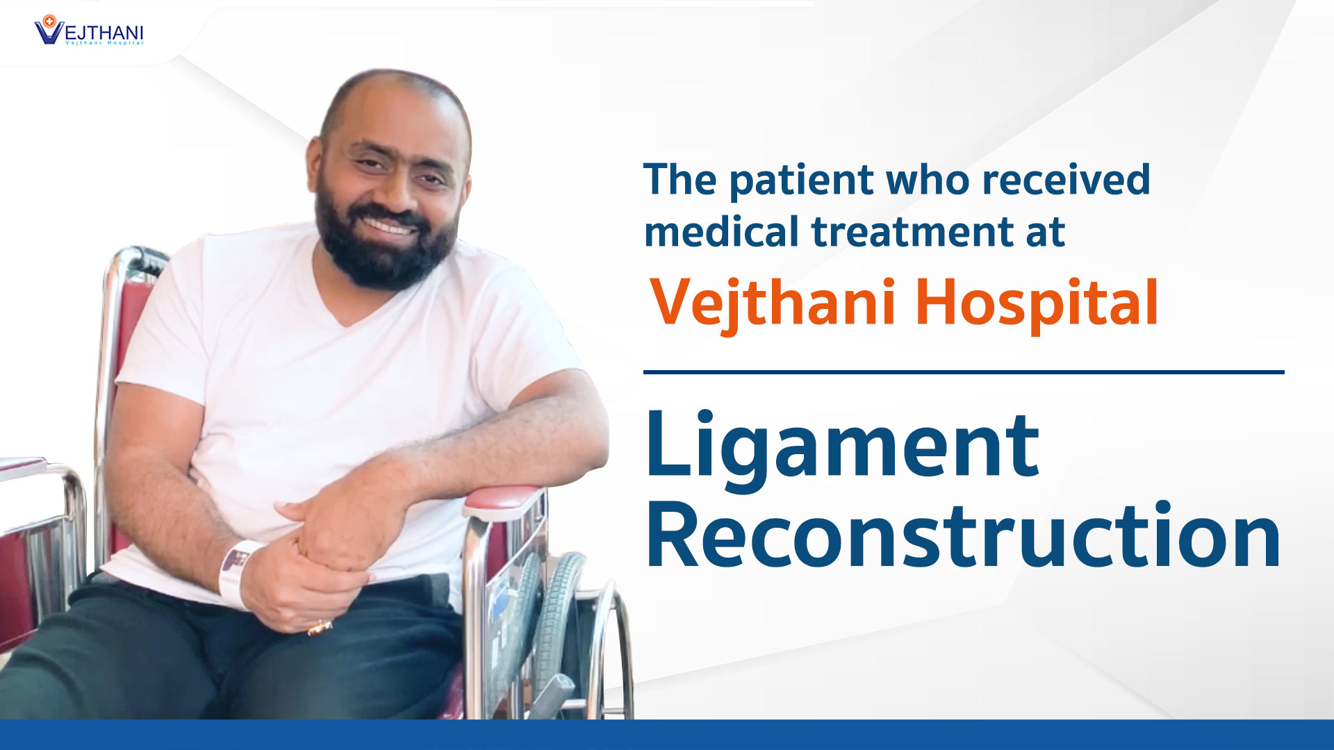
 Unusual Menstrual Symptoms Reveal Ovarian Cyst: Successful Laparoscopic CystectomyMyanmar Actress Patricia’s Intraocular Lens Surgery Experience at Vejthani HospitalLigament ReconstructionA comprehensive kid’s checkup at Vejthani Hospital’s Super Kid’s Center
 - Patient Services
- Medical Departments & Centers
- Packages & Promotions
- Health Information
Health ArticlesHealth VideosNews and UpdatesCholera is Deadly but Preventable – Protect Yourself with the Right MeasuresSmartwatch Alerts: A Warning You Can’t Afford to OverlookKidney Transplants vs Dialysis: 3 Reasons to Make a Thoughtful DecisionSevere Back Pain or Fingertip Numbness May Signal Transverse Myelitis: The Silent Killer


- About Us
Fact SheetOverseas Offices & RepresentativesAccreditations and AwardsPatient StoriesUnusual Menstrual Symptoms Reveal Ovarian Cyst: Successful Laparoscopic CystectomyMyanmar Actress Patricia’s Intraocular Lens Surgery Experience at Vejthani HospitalLigament ReconstructionA comprehensive kid’s checkup at Vejthani Hospital’s Super Kid’s Center
- Patient Services
- Medical Departments & Centers
- Packages & Promotions
- Health Information
Health ArticlesHealth VideosNews and UpdatesCholera is Deadly but Preventable – Protect Yourself with the Right MeasuresSmartwatch Alerts: A Warning You Can’t Afford to OverlookKidney Transplants vs Dialysis: 3 Reasons to Make a Thoughtful DecisionSevere Back Pain or Fingertip Numbness May Signal Transverse Myelitis: The Silent Killer
Retinal Diseases
Diagnosis
For diagnostic purposes, an ophthalmologist performs a thorough eye examination to detect any abnormalities across the eye. Furthermore, the following tests may be administered to ascertain the location and extent of the disease:
- Amsler grid test. An ophthalmologist may employ an Amsler grid to assess the clarity of your central vision. You’ll be asked to observe if the grid lines appear faded, fragmented, or distorted. Pinpointing the location of any distortion on the grid helps in assessing the extent of retinal impairment. If you have macular degeneration, you might also receive instructions to use this test for self-monitoring your condition at home.
- Optical coherence tomography (OCT). This examination method is a superb tool for capturing detailed images of the retina. It aids in diagnosing conditions such as epiretinal membranes, macular holes, and macular swelling (edema). Additionally, it enables the monitoring of the progression of age-related wet macular degeneration and its response to treatment.
- Fundus autofluorescence (FAF). FAF can be employed to assess the stage of retinal diseases, including macular degeneration. It illuminates a retinal pigment known as lipofuscin, which accumulates with retinal damage or dysfunction
- Fluorescein angiography. This examination involves the administration of a dye that enhances the visibility of blood vessels in the retina under specific lighting conditions. It aids in accurately pinpointing closed, leaking, or irregular blood vessels, as well as subtle alterations in the back of the eye.
- Indocyanine green angiography. This examination employs a dye that fluoresces under infrared light exposure. The resultant images reveal both retinal blood vessels and the deeper, less visible blood vessels behind the retina, located in a tissue known as the choroid.
- Ultrasound. This examination utilizes high-frequency sound waves, known as ultrasonography, to visualize the retina and other eye structures. It can also discern specific tissue characteristics, aiding in the diagnosis and treatment of eye tumors.
- Computed tomography (CT) and magnetic resonance imaging (MRI). Occasionally, these imaging techniques are utilized to assess eye injuries or tumors.
Treatment
The primary treatment objectives aim to halt or decelerate the progression of the disease and maintain, enhance, or restore vision. Often, damage that has occurred cannot be reversed, underscoring the importance of early detection. Your eye specialist will collaborate with you to determine the most suitable treatment plan.
Treating retinal diseases can be intricate and occasionally necessitates urgent intervention. Treatment options comprise:
- Cryopexy: In this procedure, a freezing probe is applied to the outer wall of the eye to treat a retinal tear. Intense cold penetrates the eye, freezing the retina, which later scars and secures it to the eye wall.
- Pneumatic retinopexy: Air or gas injection into the eye can aid in repairing certain types of retinal detachment. This technique may be used alongside cryopexy or laser photocoagulation.
- Scleral buckling: Surgeons employ this method to repair retinal detachment by sewing a small piece of silicone material onto the outer surface of the eye (sclera), which indents the sclera, relieving tension caused by vitreous traction and reattaching the retina. This technique may be combined with other treatments.
- Vitrectomy: This procedure involves removing the gel-like fluid (vitreous) inside the eye and replacing it with air, gas, or liquid. Vitrectomy may be necessary if bleeding or inflammation obstructs the surgeon’s view of the retina and is often part of the treatment for various conditions, including retinal tear, diabetic retinopathy, macular hole, epiretinal membrane, infection, eye trauma, or retinal detachment.
- Intravitreal injections: Medications are injected into the vitreous of the eye to treat conditions such as wet macular degeneration, diabetic retinopathy, or intraocular bleeding.
- Laser treatment: Using a laser, surgeons can repair retinal tears or holes by heating small points on the retina, which creates scarring that typically binds the retina to underlying tissue. Immediate laser treatment for a new retinal tear can reduce the risk of retinal detachment.
- Scatter laser photocoagulation: This technique can shrink irregular new blood vessels in the eye, often associated with bleeding or the threat of bleeding, particularly in cases of diabetic retinopathy. However, extensive use of this treatment may lead to the loss of peripheral or night vision.
- Retinal prosthesis implantation: People with severe vision loss or blindness caused by specific inherited retinal diseases may undergo a surgical procedure to implant a small electrode chip into the retina. This chip receives input from a video camera attached to a pair of glasses and transmits visual information that the impaired retina cannot process.