- About Us
Fact SheetOverseas Offices & RepresentativesAccreditations and AwardsPatient Stories
 Unusual Menstrual Symptoms Reveal Ovarian Cyst: Successful Laparoscopic CystectomyMyanmar Actress Patricia’s Intraocular Lens Surgery Experience at Vejthani HospitalLigament ReconstructionA comprehensive kid’s checkup at Vejthani Hospital’s Super Kid’s Center
 - Patient Services
- Medical Departments & Centers
- Packages & Promotions
- Health Information
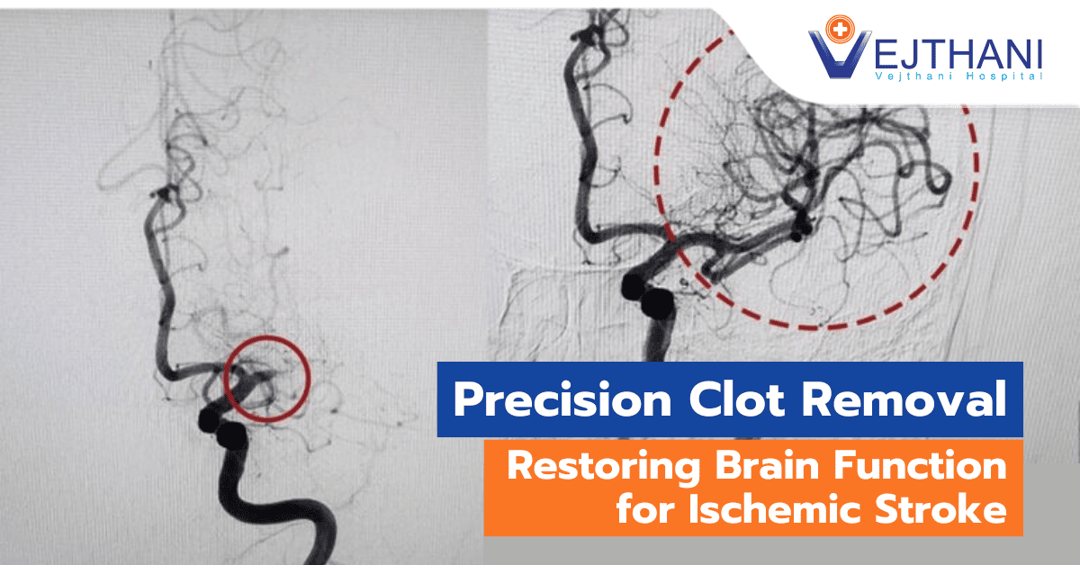
Health ArticlesHealth VideosNews and UpdatesDifferences Between Traditional Hemodialysis and Online HDFSudden Cardiac Death: A Silent Threat to AthletesLiving Donor Kidney Transplant: An Alternative Approach to Restore Renal Function without DialysisPrecision Clot Removal to Restore Brain Function in Ischemic Stroke


- About Us
Fact SheetOverseas Offices & RepresentativesAccreditations and AwardsPatient StoriesUnusual Menstrual Symptoms Reveal Ovarian Cyst: Successful Laparoscopic CystectomyMyanmar Actress Patricia’s Intraocular Lens Surgery Experience at Vejthani HospitalLigament ReconstructionA comprehensive kid’s checkup at Vejthani Hospital’s Super Kid’s Center
- Patient Services
- Medical Departments & Centers
- Packages & Promotions
- Health Information
Health ArticlesHealth VideosNews and UpdatesDifferences Between Traditional Hemodialysis and Online HDFSudden Cardiac Death: A Silent Threat to AthletesLiving Donor Kidney Transplant: An Alternative Approach to Restore Renal Function without DialysisPrecision Clot Removal to Restore Brain Function in Ischemic Stroke
Subarachnoid hemorrhage
Diagnosis
To diagnose a subarachnoid hemorrhage, your doctor is likely to suggest:
- Computed Tomography (CT) scan. This imaging test is capable of detecting bleeding in the brain. While a CT scan is highly effective when conducted correctly, it may not detect the bleed if you have a low red blood cell count (anemia) and only a small amount of blood is lost during the bleed. Your doctor may administer a contrast dye to obtain a clearer view of your blood vessels (CT angiogram).
- Magnetic Resonance Imaging (MRI). Brain hemorrhage can also be identified by this imaging technique. For an MR angiography, which shows the arteries and veins in more detail and highlights blood flow, your doctor may inject a dye into a blood vessel. In rare circumstances, when the signs are not visible on a CT scan, this may exhibit signs of a subarachnoid hemorrhage.
- Cerebral angiography. A catheter—a long, thin tube—is threaded into an artery and inserted into your brain by your healthcare professional. To make your brain’s blood vessels visible on X–ray imaging, dye is injected into them. For more precise imaging, your doctor may suggest cerebral angiography. If a subarachnoid hemorrhage is suspected but the cause is unclear or does not show up on other imaging, they might also advise the test. If the initial cerebral angiography did not reveal an aneurysm but your doctor believes one is likely, you may require a second one.
In certain cases of aneurysmal subarachnoid hemorrhages, the bleeding may not be visible on initial imaging. If your first CT scan doesn’t reveal any bleeding, your doctor might suggest a lumbar puncture. This involves inserting a needle into the lower back to withdraw a small amount of the fluid surrounding the brain and spinal cord (cerebrospinal fluid). The fluid is then examined for the presence of blood, which could indicate a subarachnoid hemorrhage.
Treatment
Treatment aims to stabilize your condition, address any existing aneurysm, and prevent complications.
Your provider monitors your breathing, blood pressure, and blood flow.
If your bleeding is due to a ruptured brain aneurysm, your provider may suggest:
- Surgery. The surgeon cuts into the scalp to access the brain aneurysm. A metal clip is then placed on the aneurysm to halt the blood flow to it.
- Endovascular embolization. The surgeon inserts a catheter into an artery and directs it to your brain. Through the catheter, detachable platinum coils are guided and placed in the aneurysm. These coils fill the aneurysm, reducing blood flow into the sac and prompting clotting. Various types of coils have been developed to treat different aneurysms.
- Other endovascular treatments. Some aneurysms can be treated with endovascular embolization, which utilizes newer technologies such as stent–assisted or balloon–assisted coiling, or devices designed to divert blood flow
Preventing complications following a subarachnoid hemorrhage is essential due to the risk of recurrent bleeding, reduced blood flow to the brain, electrolyte imbalances like low sodium, excessive fluid buildup in the brain, or blood sugar fluctuations. Nimodipine is commonly used to improve cerebral circulation after a hemorrhage.
A frequent complication of aneurysmal subarachnoid hemorrhage is delayed cerebral vasospasm, which can potentially lead to a stroke by diminishing blood flow. To prevent this, intravenous medications might be administered to increase blood pressure, or drugs that widen the brain’s blood vessels may be used.
Hydrocephalus, another frequent complication, involves fluid accumulation in the brain’s spaces and can be managed with the placement of drains either in the head (ventriculostomy catheter) or in the lower back (lumbar drains).
Repeated procedures might be necessary in some cases. Regular check–ups with your doctor are crucial to monitor any changes. Recovery may also involve physical, occupational, and speech therapies to aid rehabilitation.