Transient Ischemic Attack (TIA)
Diagnosis
In order to determine the origin of the TIA and the best course of treatment, a quick assessment of the symptoms is essential. A healthcare provider may utilize the following information to help identify the TIA’s cause and gauge the patient’s risk of having a stroke:
- Physical exam and tests. The healthcare provider will conduct both a neurological and physical examination. The vision, eye movements, speech and language, strength, reflexes, and sensory system will all be assessed by the healthcare provider. They might listen to the carotid artery in the neck using a stethoscope. A whooshing sound, or bruise, could be a sign of atherosclerosis. Alternatively, a healthcare provider might use an ophthalmoscope to search the retina’s tiny blood veins at the back of the eye for cholesterol or platelet fragments, or emboli.
The healthcare provider may perform an examination for risk factors associated with stroke, such as high blood pressure, high cholesterol, diabetes, and in certain situations, elevated homocysteine levels, among others. - Carotid ultrasonography: A carotid ultrasound may be recommended if the healthcare provider believes that the TIA may have been caused by the carotid artery. A transducer, which resembles a wand, inserts high-frequency sound waves into the neck. A healthcare provider can examine images on a screen to check for carotid artery constriction or clotting after sound waves travel through tissue and return.
- Computerized tomography (CT) or computerized tomography angiography (CTA): X-ray beams are used in CT scanning of the head to analyze the arteries in the neck and brain or to create a composite 3D image of the brain. Similar to a traditional CT scan, CTA scanning involves X-rays but also the possible injection of a contrast substance into a blood artery. A CTA scan, in contrast to a carotid ultrasonography, may assess blood arteries in the head and neck.
- Magnetic resonance imaging (MRI) or magnetic resonance angiography (MRA): By using a powerful magnetic field, these procedures are able to provide a three-dimensional composite image of the brain. While MRA may include injecting a contrast material into a blood vessel, it uses technology akin to MRI to assess the arteries in the neck and brain.
- Echocardiography: A healthcare provider may decide to conduct a transthoracic echocardiogram (TTE), a type of conventional echocardiography. During a transducer therapy, an instrument known as a transducer is moved across the chest. A transesophageal echocardiogram (TEE), a distinct kind of echocardiography, may be performed by a healthcare provider in place of the transducer, which produces an ultrasound image by reflecting sound waves off various areas of the heart. A flexible probe with an integrated transducer is inserted into the esophagus, the tube that connects the stomach to the back of the mouth, during a transesophageal echocardiogram (TEE).
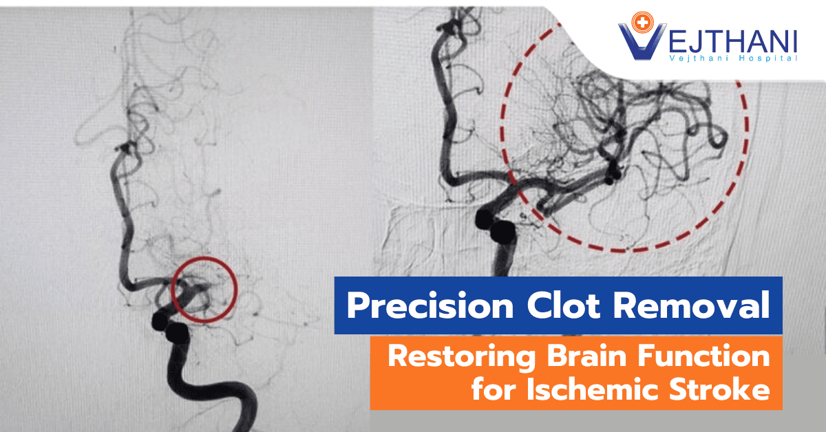
Ultrasound pictures can be produced with greater clarity and detail since the esophagus is situated immediately behind the heart. This makes certain objects, such blood clots, easier to identify that might not be apparent during a conventional echocardiogram examination. - Arteriography: This technique provides an image of the brain’s arteries that is not often visible with X-ray imaging. Through a small incision, usually in the groin, a radiologist inserts a thin, flexible tube called a catheter.
Manipulating the catheter into the carotid or vertebral artery requires passing it through the major arteries. To produce X-ray images of the brain’s arteries, the radiologist then inserts a dye through the catheter. In some situations, this process might be applied.
Treatment
Upon identifying the underlying cause of a TIA, the primary objective of treatment is to effectively address the issue and prevent the occurrence of a full-fledged stroke. Depending on the specific cause of the TIA, healthcare providers may recommend surgical interventions or balloon procedures such as angioplasty. Alternatively, they may prescribe medications aimed at reducing the risk of blood clot formation. The choice of treatment approach is tailored to the individual circumstances and contributing factors of the TIA.
- Medications: Following a TIA, healthcare providers use a variety of medications to reduce the risk of a stroke. The location, etiology, severity, and kind of TIA all influence the treatment that is chosen. A healthcare provider might recommend:
- Blood pressure medication: They lessen internal blood vessel pressure and strain. ACE inhibitors, angiotensin II receptor blockers (ARBs), calcium channel blockers, diuretics, and other medications are frequently used to treat this.
- Anti-platelet drugs: One of the circulating blood cell types, platelets, are less likely to clump together when taking these medications. Sticky platelets that have been wounded in blood arteries start to coagulate, and blood plasma proteins finish the clotting process.
Aspirin is the anti-platelet medication that is most commonly utilized. The cheapest priced treatment with the fewest possible side effects is aspirin. Aspirin can be substituted with the anti-platelet medication clopidogrel (Plavix). - As an alternative, to lower the risk of recurrent stroke, a healthcare provider may prescribe aspirin plus ticagrelor (Brilinta) for 30 days. If they want to lower blood clotting, they might prescribe Aggrenox, which is a combination of low-dose aspirin and the anti-platelet medication dipyridamole. Dipyridamole functions somewhat differently from aspirin.
- Anticoagulants: These medications inhibit the clotting of blood, hence reducing the possibility of a clot developing and becoming lodged in a cerebral blood vessel. Heparin and warfarin (Jantoven) are examples of these medications. The effect they have is on clotting-system proteins rather than platelet activity. The treatment of transient ischemic attacks (TIAs) seldom involves the short-term administration of heparin.
These medications need to be monitored closely. The healthcare provider may recommend a direct oral anticoagulant, which may be less risky than warfarin, if atrial fibrillation is detected. Examples of these medications include apixaban (Eliquis), rivaroxaban (Xarelto), edoxaban (Savaysa), or dabigatran (Pradaxa). - Statins: Medication that lowers cholesterol is called a statin. In general, they lower blood levels of low-density lipoprotein (LDL) cholesterol. That is the type of cholesterol that can accumulate inside blood vessels, producing atherosclerosis and blood vessel narrowing.
- Angioplasty: Carotid angioplasty, also known as stenting, is a technique that can be considered in certain situations. In order to keep a blocked artery open, a tiny wire tube known as a stent is inserted into the artery after it has been opened using a balloon-like device.
- Surgery: The healthcare provider may recommend a carotid endarterectomy if the patient has a moderately or severely narrowed neck (carotid) artery. By removing atherosclerotic plaques from the carotid arteries, this prophylactic procedure stops another TIA or stroke from happening. The artery is opened by making an incision, the plaques are taken out, and the artery is shut.


